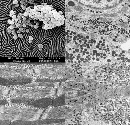
Biological Sample Preparation
SPECIMEN PREPARATION FOR TEM
- To preserve the structure of cells and tissues with minimal alteration from the living state.
- To protect them against alterations during embedding and sectioning.
- To prepare them for subsequent treatments such as staining and exposure to the electron beam.
|
Fixatives and stains |
Cellular constituents |
||||
|
Fixatives Aldehydes Uranyl acetate |
Proteins |
Nucleic acids |
Lipids |
Polysaccharides |
Phospholipids |
|
++ |
- |
- |
- |
- |
|
|
+* |
- |
++** |
- |
++ |
|
|
++ |
Destroyed |
+ |
+ (glycogen) |
++ |
|
|
|
|||||
|
++ |
- |
- |
- |
- |
|
|
++ |
++ |
- |
- |
- |
|
|
++ |
++ |
+ |
+ (glycogen) |
++ |
|
|
++ |
- |
- |
- |
- |
|
4 Procedure for fixation
4.1 Primary Fixation
The most commonly used primary fixative is 2.5% glutaraldehyde in 0.1M sodium phosphate buffer (PB; pH 7.4). For certain tissues, a mixture of 4% paraformaldehyde and 1% glutaraldehyde in 0.1 M PB (pH 7.4) is used and this is called Karnovsky's fixative.
- Fixation through immersion
The whole organ or parts of it is cut and put directly in the fixative. Due to slow penetration of the fixative, the ideal size of the tissue should be about 1-2 mm2 thick. Depending upon the type of tissues, fixation is carried out for 2-12 hours at 4oC.
- Fixation through perfusion
When organs are large or dissection is difficult, fixation is carried out by perfusing the intact organism with fixative. This is carried out by injecting fixative through the heart, using transfusion set (bottle with the polythene catheter containing the fixative). For an adult rat, about 300 to 400 ml of the fixative is perfused. The various steps are
- The animals are anesthetised with sodium pentobarbitone (30mg / kg body weight)
- The heart is exposed.
- The ascending aorta is canulated with a catheter of 1 mm diameter through the left ventricle.
- The right auricle is incised for the outflow of the fixative.
- The fixative is allowed to flow rapidly for a minute and then the flow is reduced to about 6 ml/minute.
- Proper fixation is adjudged by the hardening of organs and stiffening of limbs.
After perfusion, organs are dissected out, the tissues are cut into 1-2 mm2 size and fixed in the same fixative for 2-4 hours at 4oC.
Washing
After fixation, specimens are rinsed thoroughly in 0.1 M PB to wash off the excess fixative. If traces of fixatives are left in the specimen, they may react with OsO4, producing precipitate of reduced osmium in the specimen.
4.2 Post-fixation (Secondary fixation)
Osmium tetroxide (1% solution) is most frequently used as a secondary fixative. It is used after glutaraldehyde fixation. It reacts mainly with lipids, converting unsaturated fatty acids to stable glycol osmate. It also acts as an electron dense stain.
Washing: After fixation, specimens are rinsed thoroughly in buffer to wash off excess fixative.
4.3 Dehydration
Biological tissues contain about 75-80% water. It is removed before tissue embedding in water immiscible resins. Tissue water is removed by passing the tissues through a series of ascending concentrations of the dehydrating agent. Ethanol or acetone is used as a dehydrating agent. Acetone has an advantage over ethanol, as it is more readily miscible with clearing agents, viz. xylene and toluene.
4.4 Clearing
Clearing is a process of replacement of the dehydrating agent with a solvent that facilitates penetration of resin into tissues. Though acetone is easily miscible with resin, it is useful to clear it off with a clearing agent to facilitate penetration of resin. Xylene, toluene and epoxy propane are good clearing agents.
4.5 Infiltration
Very few tissues are rigid enough to be cut into thin sections without an additional support. Therefore, tissues must be infiltrated and embedded into a resin and made into hard blocks that allow thin sectioning easily. Infiltration is done by gradually decreasing the concentration of the clearing agent and increasing the concentration of the embedding medium. In case of hard and plant tissues, slow diffusion of the embedding medium is a problem. This can be overcome by carrying out infiltration under vacuum. Infiltration is carried out with liquid resins. Resins are of two kinds, as follows:
4.6 Kinds of resin
- Epoxy resins (water immiscible resin): Araldite CY212, Epon 812, ERL 4206
• Acrylic resins (water miscible resins)
Durcapan, hydroxypropyl methacrylate, LR gold and LR white, Lowicryl resins
4.7 Embedding
Embedding is done using flat molds or plastic capsules. After filling the molds/capsules with the embedding medium, tissue is placed. In capsules, tissue is settled at the bottom and it is filled with embedding medium. Paper label written with pencil is put along the sidewall of the capsule or inside the mold. When a specific orientation is required, flat embedding mold is used.
4.8 Polymerization
It is the process by which resin is allowed to harden by heat treatment. At 50oC, the embedding medium becomes less viscous which facilitates penetration into the tissue. So the specimen is treated first at 50o C for overnight, then at 60oC, until the resin gets hardened or polymerized (takes about 48-72 hours).

